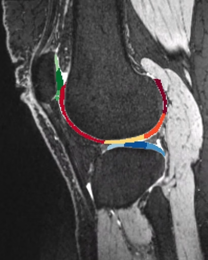
Musculoskeletal disorders and injuries are among the most common health issues affecting millions of people, significantly impacting their quality of life. These conditions can affect your muscles, bones, and joints. At Medicana Winchester, our advanced 3T MRI imaging plays a crucial role in the diagnosis, treatment, and prevention of these conditions by producing detailed images of the musculoskeletal system. The higher magnetic field strength of our 3T MRI provides exceptional image quality and greater detail compared to traditional MRI scanners.
A musculoskeletal (MSK) MRI is a non-invasive imaging technique that uses strong magnetic fields and radio waves to produce detailed images of the bones, muscles, tendons, ligaments, cartilage, and other connective tissues. Unlike X-rays or CT scans, which use ionising radiation, MRI scans are a safer option for repeated use as they rely on magnetic fields.
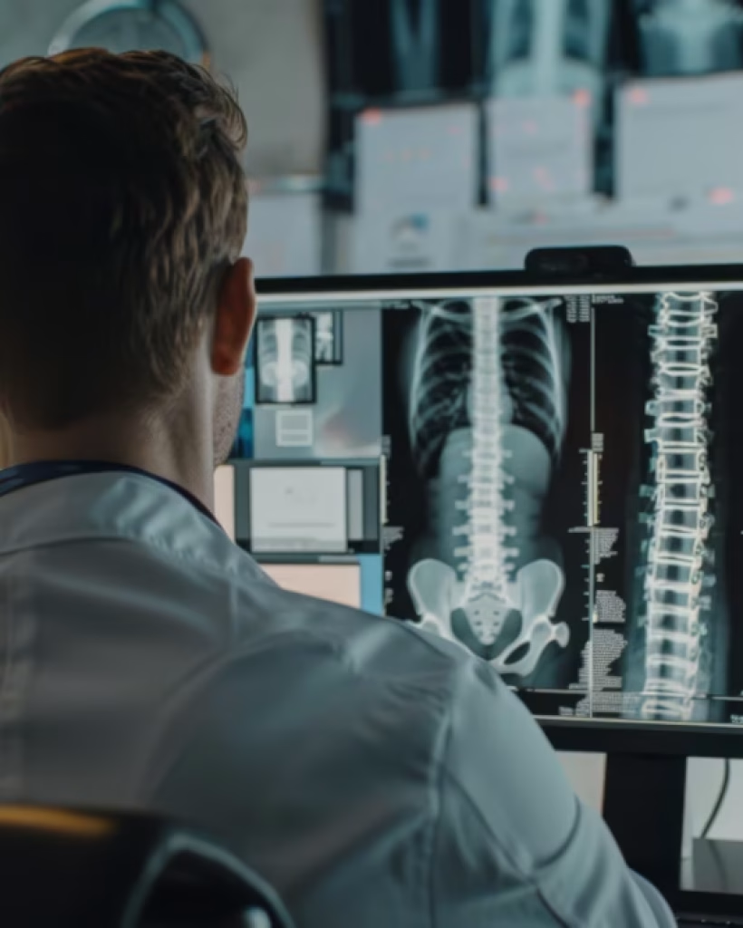
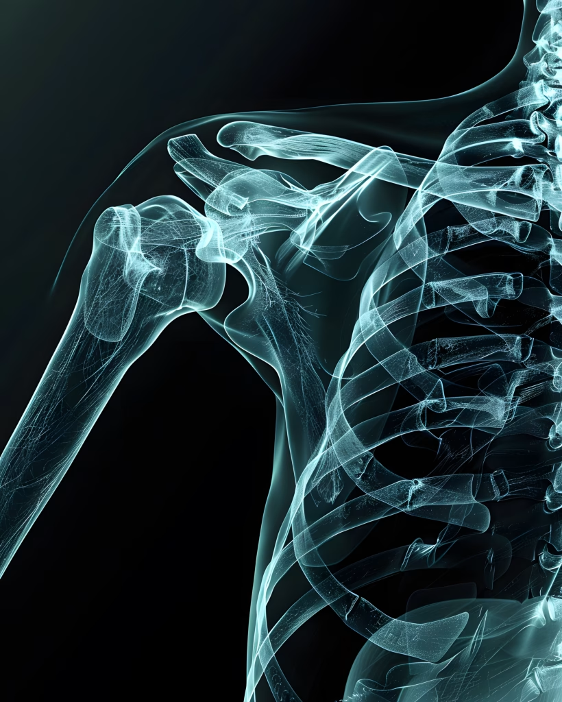
The gain in signal-to-noise ratio (SNR) afforded by 3T MRI systems has tremendous clinical applications in the musculoskeletal system. This technology offers a significant potential for improving diagnostic abilities and patient care. Key benefits include:
Improved Image Quality: The higher magnetic field strength and greater signal-to-noise ratio provide superior image quality, which is particularly helpful in assessing small structures and soft tissues in children.
Greater Diagnostic Accuracy: The enhanced resolution allows for greater accuracy in diagnosing injuries to articular cartilage, glenoid labrum of the shoulder, intrinsic ligaments of the wrist, and collateral ligaments of the elbow and ankle, which are often evaluated suboptimally on lower-strength systems.
Faster Scan Times: 3T MRIs provide higher detailed images in less time, making the scan more comfortable for the patient.
Detailed Joint Assessment: The higher quality scans can detect smaller abnormalities or fractures of joints and even show bleeding involved with fractures. They also offer detailed imaging of ligaments, tendons, and cartilage to assess joint stability.
Musculoskeletal MRI scans are invaluable for diagnosing a variety of conditions, including:
Injuries: Muscle tears, ligament sprains, and tendon strains.
Arthritis: Identifying and assessing the severity of osteoarthritis and rheumatoid arthritis.
Bone Disorders: Detecting fractures, bone infections (osteomyelitis), and bone tumours.
Soft Tissue Conditions: Inflammation, tears, and other soft tissue abnormalities.
Spinal Issues: Identifying herniated discs, spinal stenosis, and other spinal problems.
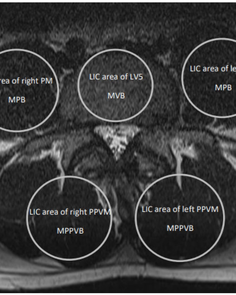
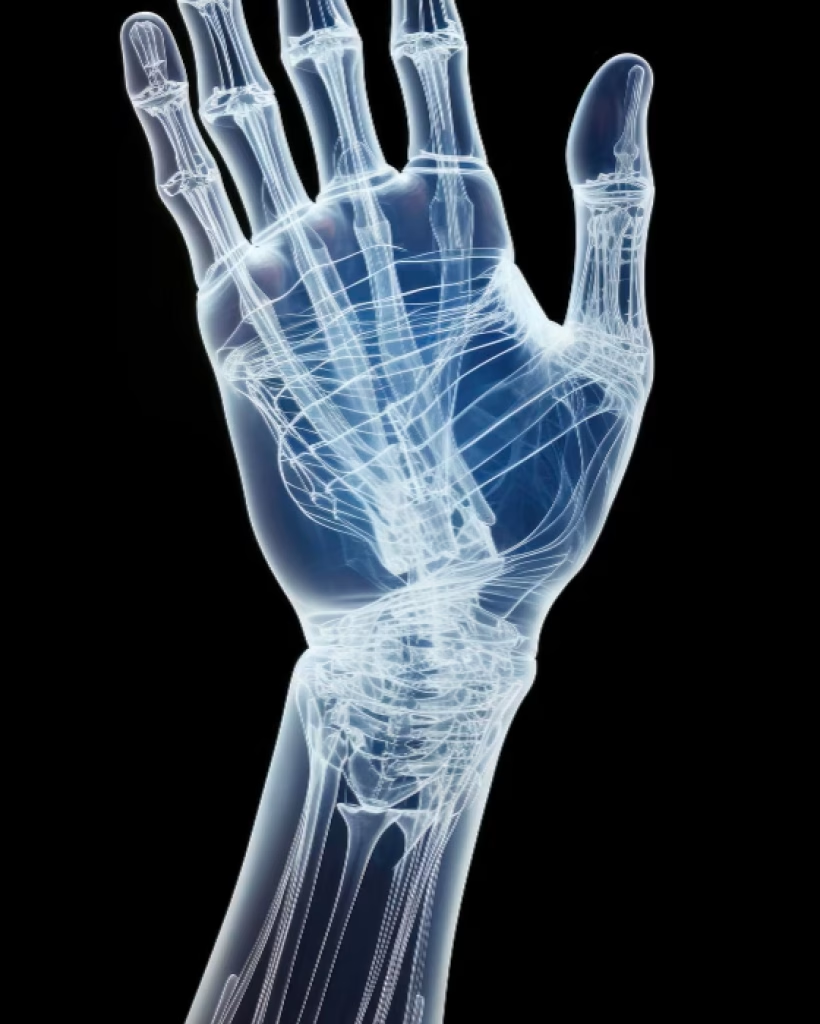
Here’s a step-by-step overview of what happens during a musculoskeletal MRI:
Preparation: You will be asked to remove all metal objects, such as jewellery and watches, as they can interfere with the magnetic field. You may also be asked to change into a hospital gown.
Positioning: You’ll lie on a moveable table that slides into the MRI machine. Our dedicated Patient Care Team will ensure you are comfortable.
During the Scan: The MRI machine produces loud tapping or thumping sounds, so you will be given earplugs or headphones. Staying still is crucial to obtain clear images.
Contrast Agents: A contrast agent (gadolinium) may sometimes be injected to enhance the images, which helps highlight specific tissues and abnormalities.
Duration: The scan usually takes between 30-60 minutes, depending on the complexity of the examination.
Please click the button below to request your private medical consultation.
Quick, secure, and personalised appointment requests in just a few steps.
To learn more about arranging your self-pay healthcare at Medicana Winchester Clinic, please don’t hesitate to contact us:
📞 Call us: +44 1962 587821
Stay informed about the latest health news and exclusive offers. Subscribe to our newsletter today!